Iron »
PDB 7oqy-7p7j »
7p17 »
Iron in PDB 7p17: F(M197)H Mutant Structure of Photosynthetic Reaction Center From Rhodobacter Sphaeroides Strain Rv By Fixed-Target Serial Synchrotron Crystallography (Room Temperature, 12KEV)
Protein crystallography data
The structure of F(M197)H Mutant Structure of Photosynthetic Reaction Center From Rhodobacter Sphaeroides Strain Rv By Fixed-Target Serial Synchrotron Crystallography (Room Temperature, 12KEV), PDB code: 7p17
was solved by
A.G.Gabdulkhakov,
G.K.Selikhanov,
S.Guenther,
A.Meents,
T.Y.Fufina,
L.G.Vasilieva,
with X-Ray Crystallography technique. A brief refinement statistics is given in the table below:
| Resolution Low / High (Å) | 47.05 / 2.22 |
| Space group | P 42 21 2 |
| Cell size a, b, c (Å), α, β, γ (°) | 102.5, 102.5, 237.4, 90, 90, 90 |
| R / Rfree (%) | 17.7 / 21.5 |
Other elements in 7p17:
The structure of F(M197)H Mutant Structure of Photosynthetic Reaction Center From Rhodobacter Sphaeroides Strain Rv By Fixed-Target Serial Synchrotron Crystallography (Room Temperature, 12KEV) also contains other interesting chemical elements:
| Magnesium | (Mg) | 4 atoms |
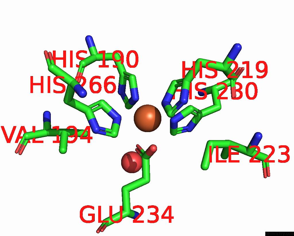
Iron Binding Sites:
The binding sites of Iron atom in the F(M197)H Mutant Structure of Photosynthetic Reaction Center From Rhodobacter Sphaeroides Strain Rv By Fixed-Target Serial Synchrotron Crystallography (Room Temperature, 12KEV)
(pdb code 7p17). This binding sites where shown within
5.0 Angstroms radius around Iron atom.
In total only one binding site of Iron was determined in the F(M197)H Mutant Structure of Photosynthetic Reaction Center From Rhodobacter Sphaeroides Strain Rv By Fixed-Target Serial Synchrotron Crystallography (Room Temperature, 12KEV), PDB code: 7p17:
In total only one binding site of Iron was determined in the F(M197)H Mutant Structure of Photosynthetic Reaction Center From Rhodobacter Sphaeroides Strain Rv By Fixed-Target Serial Synchrotron Crystallography (Room Temperature, 12KEV), PDB code: 7p17:
Iron binding site 1 out of 1 in 7p17
Go back to
Iron binding site 1 out
of 1 in the F(M197)H Mutant Structure of Photosynthetic Reaction Center From Rhodobacter Sphaeroides Strain Rv By Fixed-Target Serial Synchrotron Crystallography (Room Temperature, 12KEV)
Mono view
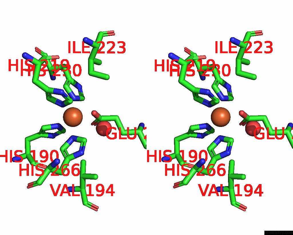
Stereo pair view
Mono view
Stereo pair view
A full contact list of Iron with other atoms in the Fe binding
site number 1 of F(M197)H Mutant Structure of Photosynthetic Reaction Center From Rhodobacter Sphaeroides Strain Rv By Fixed-Target Serial Synchrotron Crystallography (Room Temperature, 12KEV) within 5.0Å range:
|
Reference:
A.G.Gabdulkhakov,
G.K.Selikhanov,
S.Guenther,
A.Meents,
T.Y.Fufina,
L.G.Vasilieva.
X-Ray Structure of Rhodobacter Sphaeroides Reaction Center with M197 Phe-His Substitution Clarifies Properties of the Mutant Complex To Be Published.
Page generated: Thu Aug 8 14:53:48 2024
Last articles
Fe in 2BCCFe in 2BH4
Fe in 2BGV
Fe in 2BC5
Fe in 2BDM
Fe in 2BCN
Fe in 2B7S
Fe in 2B7R
Fe in 2B7H
Fe in 2B3Y