Iron »
PDB 8iba-8irf »
8iim »
Iron in PDB 8iim: H109K Mutant of Uracil Dna Glycosylase X
Enzymatic activity of H109K Mutant of Uracil Dna Glycosylase X
All present enzymatic activity of H109K Mutant of Uracil Dna Glycosylase X:
3.2.2.27;
3.2.2.27;
Protein crystallography data
The structure of H109K Mutant of Uracil Dna Glycosylase X, PDB code: 8iim
was solved by
S.Aroli,
with X-Ray Crystallography technique. A brief refinement statistics is given in the table below:
| Resolution Low / High (Å) | 26.41 / 1.60 |
| Space group | P 1 21 1 |
| Cell size a, b, c (Å), α, β, γ (°) | 36.45, 51.75, 54.63, 90, 104.83, 90 |
| R / Rfree (%) | 16.4 / 18.2 |
Iron Binding Sites:
The binding sites of Iron atom in the H109K Mutant of Uracil Dna Glycosylase X
(pdb code 8iim). This binding sites where shown within
5.0 Angstroms radius around Iron atom.
In total 4 binding sites of Iron where determined in the H109K Mutant of Uracil Dna Glycosylase X, PDB code: 8iim:
Jump to Iron binding site number: 1; 2; 3; 4;
In total 4 binding sites of Iron where determined in the H109K Mutant of Uracil Dna Glycosylase X, PDB code: 8iim:
Jump to Iron binding site number: 1; 2; 3; 4;
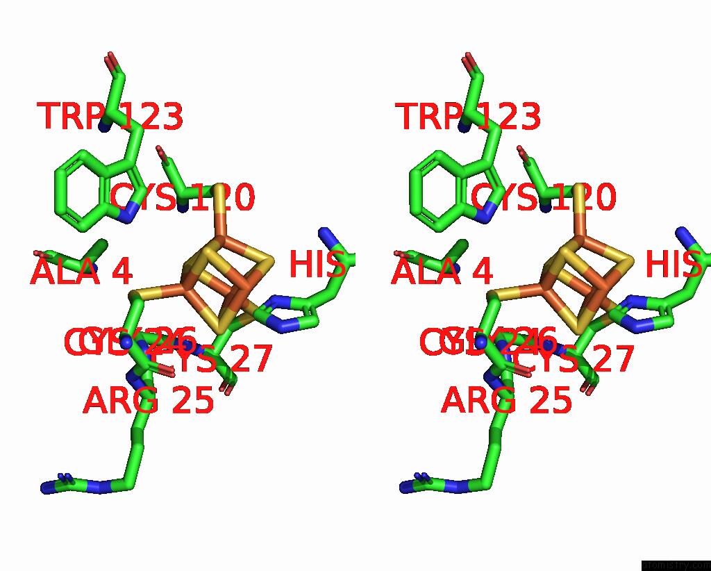
Iron binding site 1 out of 4 in 8iim
Go back to
Iron binding site 1 out
of 4 in the H109K Mutant of Uracil Dna Glycosylase X
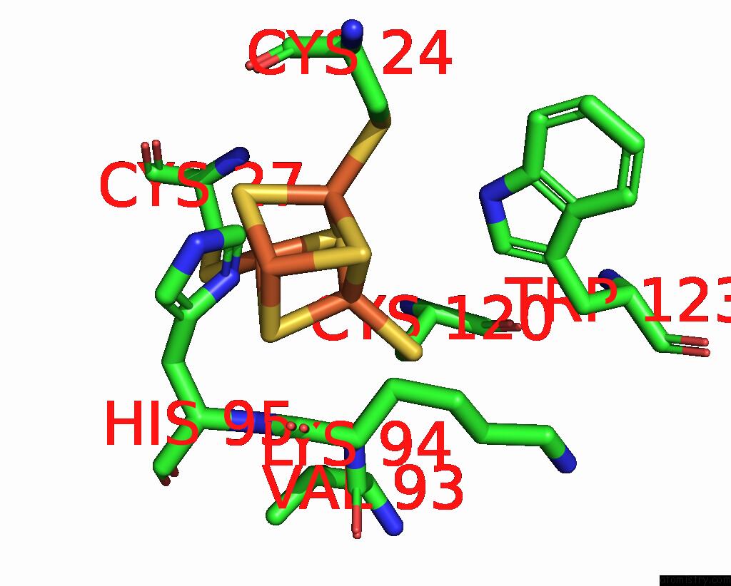
Mono view
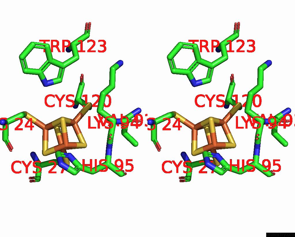
Stereo pair view
Mono view
Stereo pair view
A full contact list of Iron with other atoms in the Fe binding
site number 1 of H109K Mutant of Uracil Dna Glycosylase X within 5.0Å range:
|
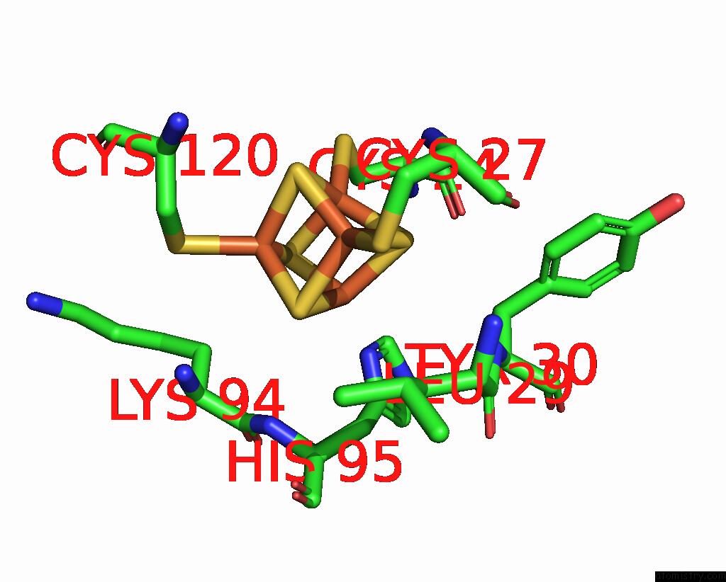
Iron binding site 2 out of 4 in 8iim
Go back to
Iron binding site 2 out
of 4 in the H109K Mutant of Uracil Dna Glycosylase X

Mono view
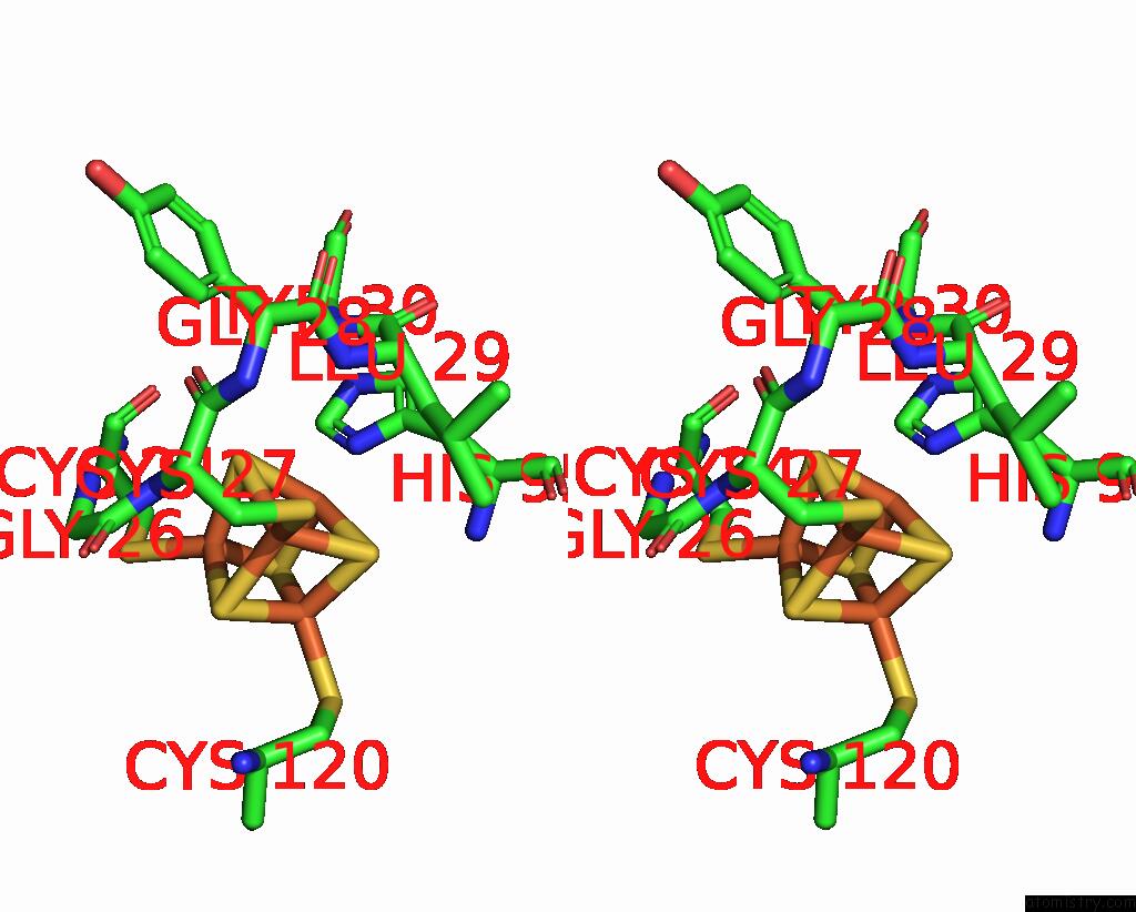
Stereo pair view
Mono view
Stereo pair view
A full contact list of Iron with other atoms in the Fe binding
site number 2 of H109K Mutant of Uracil Dna Glycosylase X within 5.0Å range:
|
Iron binding site 3 out of 4 in 8iim
Go back to
Iron binding site 3 out
of 4 in the H109K Mutant of Uracil Dna Glycosylase X
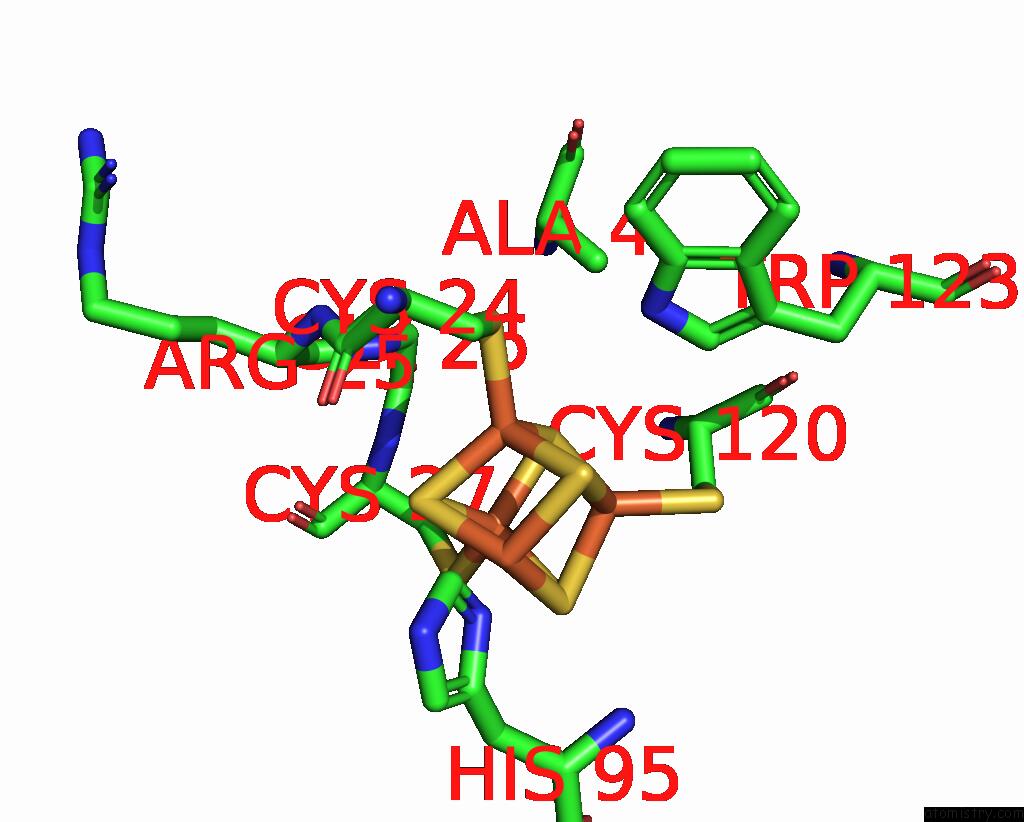
Mono view
Stereo pair view
Mono view
Stereo pair view
A full contact list of Iron with other atoms in the Fe binding
site number 3 of H109K Mutant of Uracil Dna Glycosylase X within 5.0Å range:
|
Iron binding site 4 out of 4 in 8iim
Go back to
Iron binding site 4 out
of 4 in the H109K Mutant of Uracil Dna Glycosylase X
Mono view

Stereo pair view
Mono view
Stereo pair view
A full contact list of Iron with other atoms in the Fe binding
site number 4 of H109K Mutant of Uracil Dna Glycosylase X within 5.0Å range:
|
Reference:
S.Aroli,
E.J.Woo,
B.Gopal,
U.Varshney.
Mutational and Structural Analyses of Udgx: Insights Into the Active Site Pocket Architecture and Its Evolution. Nucleic Acids Res. 2023.
ISSN: ESSN 1362-4962
PubMed: 37283083
DOI: 10.1093/NAR/GKAD486
Page generated: Thu Aug 7 18:10:34 2025
ISSN: ESSN 1362-4962
PubMed: 37283083
DOI: 10.1093/NAR/GKAD486
Last articles
Fe in 8Q1WFe in 8Q1V
Fe in 8Q1P
Fe in 8Q0O
Fe in 8Q0Q
Fe in 8Q1B
Fe in 8Q0M
Fe in 8Q0F
Fe in 8Q0A
Fe in 8Q0J