Iron »
PDB 8dju-8e3v »
8dzl »
Iron in PDB 8dzl: Structure of the K39Q Mutant of Rat Somatic Cytochrome C at 1.36A
Protein crystallography data
The structure of Structure of the K39Q Mutant of Rat Somatic Cytochrome C at 1.36A, PDB code: 8dzl
was solved by
B.F.P.Edwards,
M.Huettemann,
A.Vaishnav,
J.Brunzelle,
P.Morse,
J.Wan,
with X-Ray Crystallography technique. A brief refinement statistics is given in the table below:
| Resolution Low / High (Å) | 58.10 / 1.36 |
| Space group | P 21 21 21 |
| Cell size a, b, c (Å), α, β, γ (°) | 33.578, 61.153, 186.065, 90, 90, 90 |
| R / Rfree (%) | 14.1 / 18 |
Iron Binding Sites:
The binding sites of Iron atom in the Structure of the K39Q Mutant of Rat Somatic Cytochrome C at 1.36A
(pdb code 8dzl). This binding sites where shown within
5.0 Angstroms radius around Iron atom.
In total 4 binding sites of Iron where determined in the Structure of the K39Q Mutant of Rat Somatic Cytochrome C at 1.36A, PDB code: 8dzl:
Jump to Iron binding site number: 1; 2; 3; 4;
In total 4 binding sites of Iron where determined in the Structure of the K39Q Mutant of Rat Somatic Cytochrome C at 1.36A, PDB code: 8dzl:
Jump to Iron binding site number: 1; 2; 3; 4;
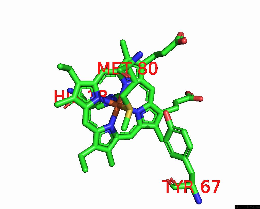
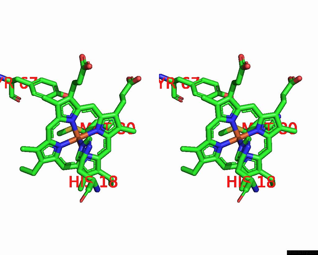
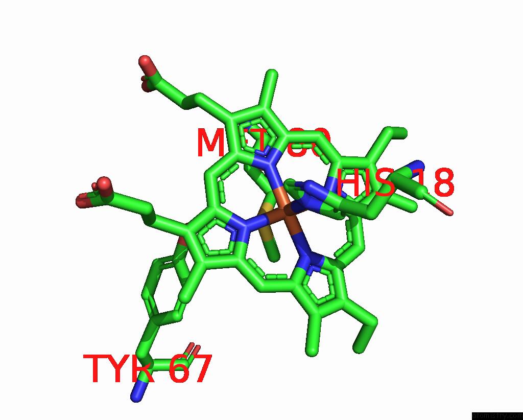
Iron binding site 1 out of 4 in 8dzl
Go back to
Iron binding site 1 out
of 4 in the Structure of the K39Q Mutant of Rat Somatic Cytochrome C at 1.36A

Mono view
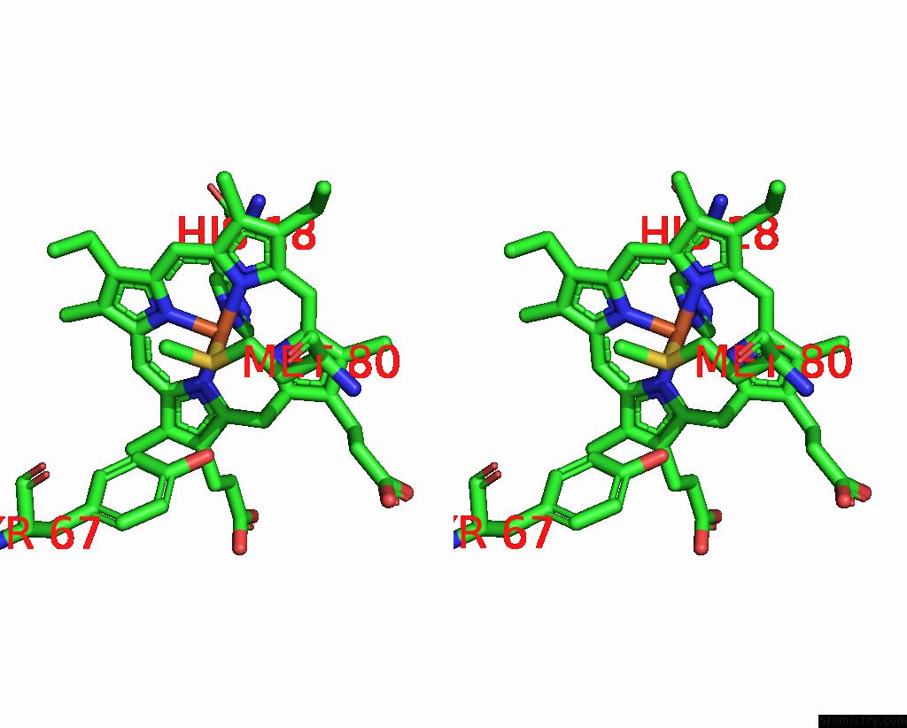
Stereo pair view
Mono view
Stereo pair view
A full contact list of Iron with other atoms in the Fe binding
site number 1 of Structure of the K39Q Mutant of Rat Somatic Cytochrome C at 1.36A within 5.0Å range:
|
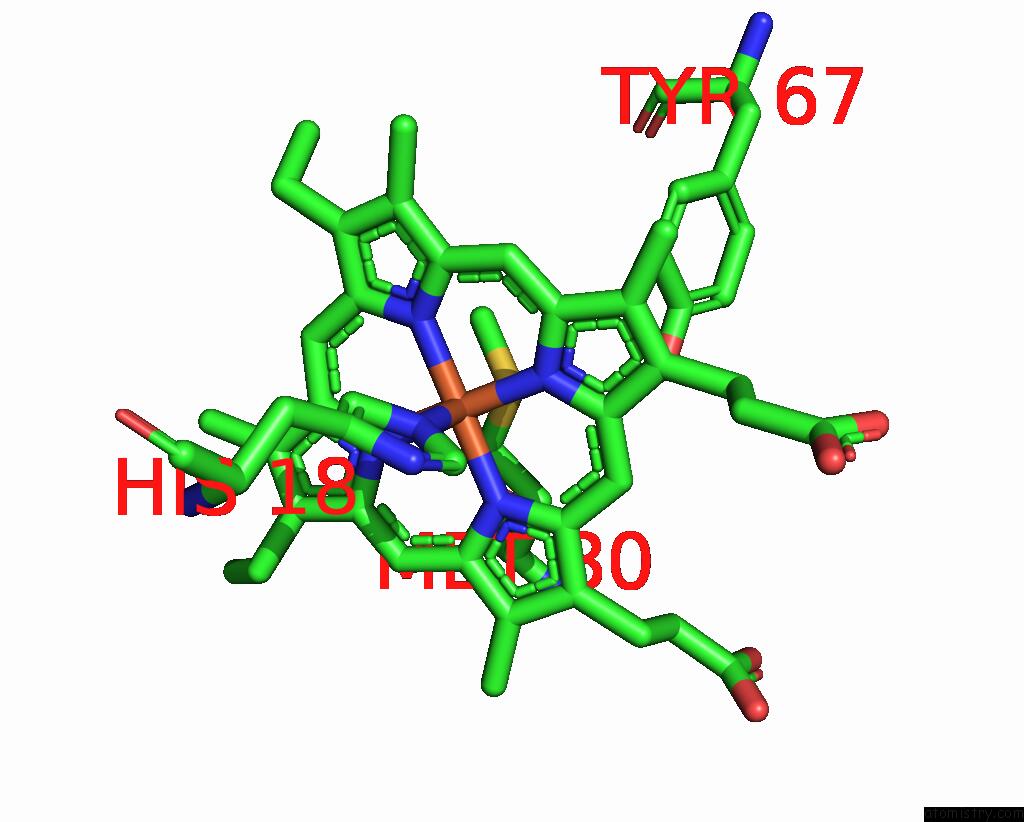
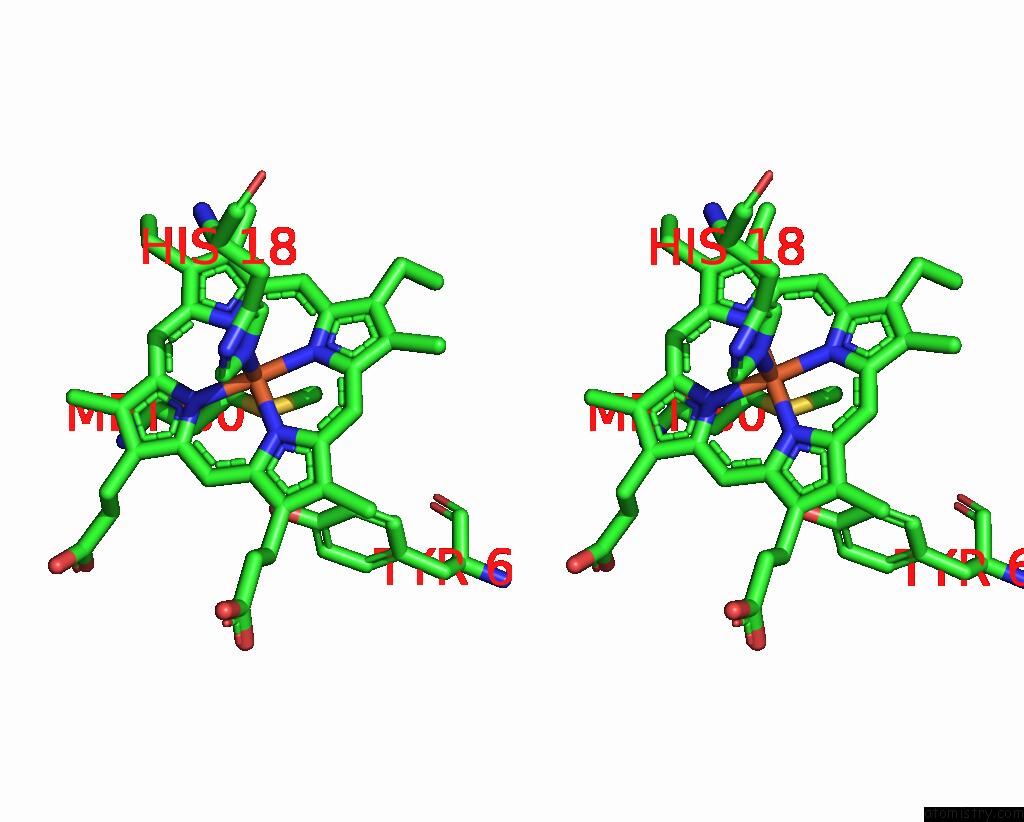
Iron binding site 2 out of 4 in 8dzl
Go back to
Iron binding site 2 out
of 4 in the Structure of the K39Q Mutant of Rat Somatic Cytochrome C at 1.36A
Mono view
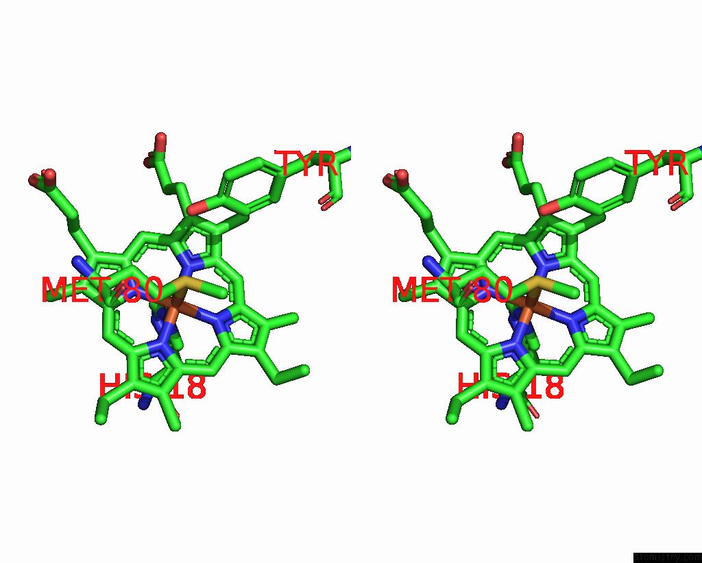
Stereo pair view
Mono view
Stereo pair view
A full contact list of Iron with other atoms in the Fe binding
site number 2 of Structure of the K39Q Mutant of Rat Somatic Cytochrome C at 1.36A within 5.0Å range:
|
Iron binding site 3 out of 4 in 8dzl
Go back to
Iron binding site 3 out
of 4 in the Structure of the K39Q Mutant of Rat Somatic Cytochrome C at 1.36A
Mono view
Stereo pair view
Mono view
Stereo pair view
A full contact list of Iron with other atoms in the Fe binding
site number 3 of Structure of the K39Q Mutant of Rat Somatic Cytochrome C at 1.36A within 5.0Å range:
|
Iron binding site 4 out of 4 in 8dzl
Go back to
Iron binding site 4 out
of 4 in the Structure of the K39Q Mutant of Rat Somatic Cytochrome C at 1.36A
Mono view
Stereo pair view
Mono view
Stereo pair view
A full contact list of Iron with other atoms in the Fe binding
site number 4 of Structure of the K39Q Mutant of Rat Somatic Cytochrome C at 1.36A within 5.0Å range:
|
Reference:
P.R.Morse,
H.Kalpage,
J.Wan,
A.Vaishnav,
D.D.Chowdhury,
J.S.Brunzelle,
B.F.P.Edwards,
M.Huettemann.
Lysine 39 Acetylation of Cytochrome C in Ischemic Skeletal Muscle Stimulates Respiration While Attenuating Cell Death Nat Commun 2023.
ISSN: ESSN 2041-1723
DOI: 10.1038/S41467-023-39820-8
Page generated: Thu Aug 7 16:01:08 2025
ISSN: ESSN 2041-1723
DOI: 10.1038/S41467-023-39820-8
Last articles
Fe in 8OLPFe in 8OLH
Fe in 8OHY
Fe in 8K9E
Fe in 8K9F
Fe in 8OGI
Fe in 8OEZ
Fe in 8OGG
Fe in 8OFL
Fe in 8OEM