Iron »
PDB 8euh-8f9n »
8f68 »
Iron in PDB 8f68: E. Coli Cytochrome BO3 Ubiquinol Oxidase Monomer
Enzymatic activity of E. Coli Cytochrome BO3 Ubiquinol Oxidase Monomer
All present enzymatic activity of E. Coli Cytochrome BO3 Ubiquinol Oxidase Monomer:
7.1.1.3;
7.1.1.3;
Other elements in 8f68:
The structure of E. Coli Cytochrome BO3 Ubiquinol Oxidase Monomer also contains other interesting chemical elements:
| Copper | (Cu) | 1 atom |
Iron Binding Sites:
The binding sites of Iron atom in the E. Coli Cytochrome BO3 Ubiquinol Oxidase Monomer
(pdb code 8f68). This binding sites where shown within
5.0 Angstroms radius around Iron atom.
In total 2 binding sites of Iron where determined in the E. Coli Cytochrome BO3 Ubiquinol Oxidase Monomer, PDB code: 8f68:
Jump to Iron binding site number: 1; 2;
In total 2 binding sites of Iron where determined in the E. Coli Cytochrome BO3 Ubiquinol Oxidase Monomer, PDB code: 8f68:
Jump to Iron binding site number: 1; 2;
Iron binding site 1 out of 2 in 8f68
Go back to
Iron binding site 1 out
of 2 in the E. Coli Cytochrome BO3 Ubiquinol Oxidase Monomer
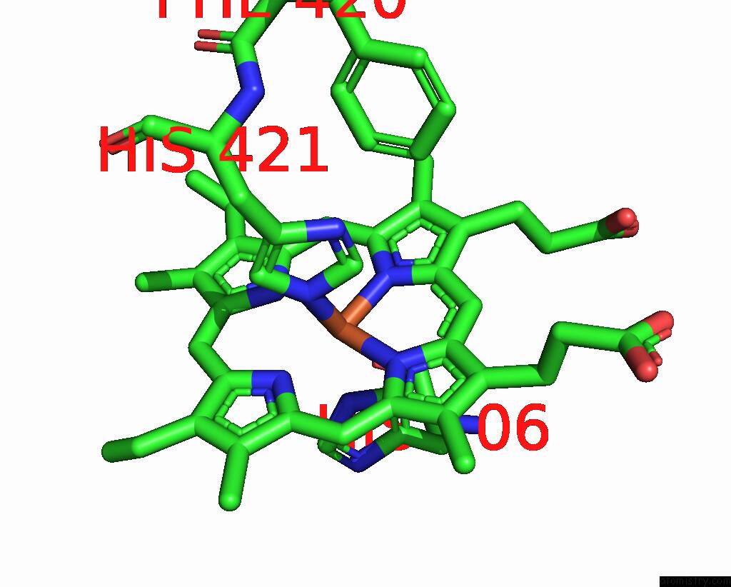
Mono view
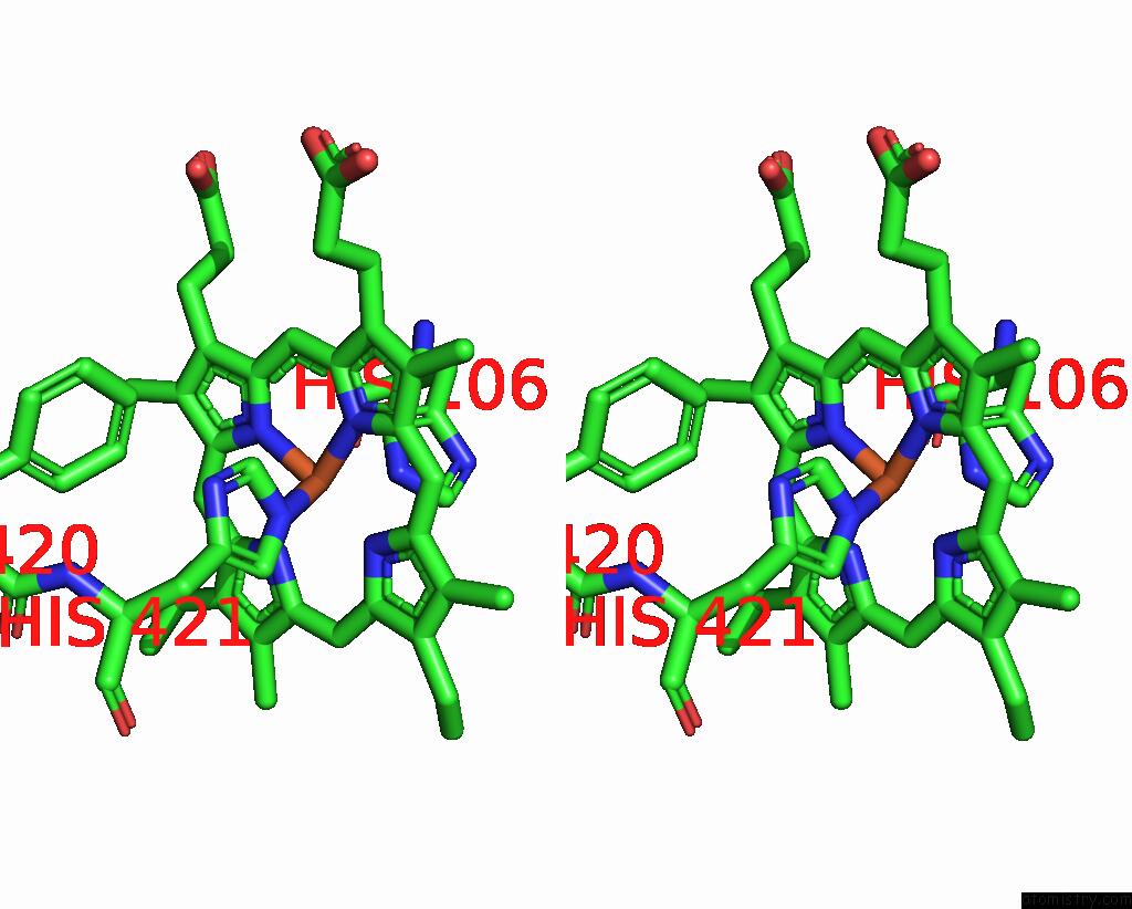
Stereo pair view
Mono view
Stereo pair view
A full contact list of Iron with other atoms in the Fe binding
site number 1 of E. Coli Cytochrome BO3 Ubiquinol Oxidase Monomer within 5.0Å range:
|
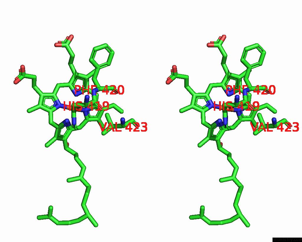
Iron binding site 2 out of 2 in 8f68
Go back to
Iron binding site 2 out
of 2 in the E. Coli Cytochrome BO3 Ubiquinol Oxidase Monomer

Mono view
Stereo pair view
Mono view
Stereo pair view
A full contact list of Iron with other atoms in the Fe binding
site number 2 of E. Coli Cytochrome BO3 Ubiquinol Oxidase Monomer within 5.0Å range:
|
Reference:
Y.Guo,
E.Karimullina,
D.Borek,
A.Savchenko.
Monomer and Dimer Structures of E. Coli Cytochrome BO3 Ubiquinol Oxidase To Be Published.
Page generated: Thu Aug 7 16:50:49 2025
Last articles
Fe in 8OLPFe in 8OLH
Fe in 8OHY
Fe in 8K9E
Fe in 8K9F
Fe in 8OGI
Fe in 8OEZ
Fe in 8OGG
Fe in 8OFL
Fe in 8OEM